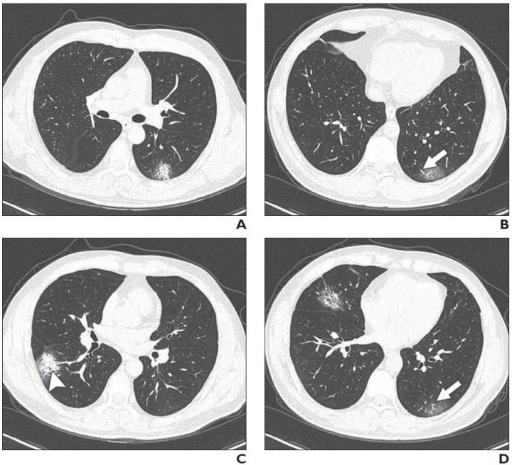
A recent study compares the chest CT scans with the clinical conditions of coronavirus disease (COVID-19) pneumonia in 101 patients. According to the findings, 86.1 percent of patients with COVID-19 pneumonia had a CT feature called ground-glass opacities (GGO), 64.4 percent had mixed GGO, and 71.3 percent had vascular enlargement in the lesion.
They described the lesions (area of tissue damage) on the CT scans as “more likely to have peripheral distribution (87.1%) and bilateral involvement (82.2%) and be lower lung predominant (54.5%) and multifocal (54.5%).”
The 101 cases were collected from four institutions in Wuhan province of China, the epicenter of the COVID-19 outbreak. They divided the cases into nonemergency (mild or common disease) and emergency (severe or fatal disease).
The patients (70.2 percent) were mostly aged 21-50 years and most (78.2 percent) had fever as the onset symptom. The patients in the emergency group were older than those in the nonemergency group. However, there were no significant different in underlying disease between the two groups.
“Architectural distortion, traction bronchiectasis, and pleural effusions, which may reflect the viral load and virulence of COVID-19, were statistically different between the two groups and may help us to identify the emergency type disease,” said the lead authors in their press release.
The findings will be published in the American Journal of Roentgenology (AJR).
